CellTiter-Glo® 3D Cell Viability Assay
A Cell Viability Assay Validated for 3D Microtissue Cultures
- Accurate 3D cytotoxicity determination
- Easy assay implementation
- Simple, 30-minute protocol


Catalog Number:
Size
Catalog Number: G9681
Catalog Number: G9682
Catalog Number: G9683
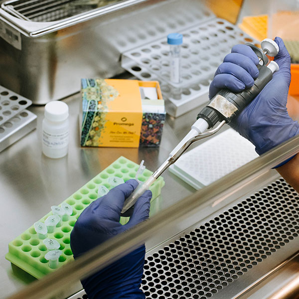
How Does CellTiter-Glo 3D Work?
The CellTiter-Glo® 3D Cell Viability Assay is designed for determining cell viability in 3D microtissue spheroids. The assay reagent penetrates large spheroids and has increased lytic capacity—allowing more accurate determination of viability compared to other assay methods.
Based on the same reliable chemistry as the classic CellTiter-Glo® Assay, this new 3D assay reagent measures ATP as an indicator of viability and generates a luminescent readout that is much more sensitive than colorimetric or fluorescence-based methods. The simple, 30-minute protocol and ready-to-use reagent allows for fast results.
Learn how the CellTiter-Glo® 3D Cell Viability Assay can be used to assess drug toxicity in organoids.
Better ATP Recovery from Larger 3D Microtissue Spheroids
| Diameter of Spheroid (μm) | Classic CellTiter-Glo® Assay (pmol/microtissue) | CellTiter-Glo® 3D Assay (pmol/microtissue) | Ratio |
|---|---|---|---|
| 188 | 16 ± 4 | 17 ± 4 | 1.10 |
| 386 | 79 ± 3 | 94 ± 11 | 1.19 |
| 459 | 103 ± 2 | 126 ± 11 | 1.22 |
| 565 | 127 ± 3 | 178 ± 17 | 1.40 |
Better Lytic Capacity
Improved 3D Microtissue Penetration, More Accurate Viability Data
HCT116 colon cancer spheroids were generated by seeding cells in the InSphero GravityPLUS™ 96-well hanging-drop platform and grown for 4 days.
Panel A. An equivalent volume of reagent was added to all samples, and after 5 minutes of shaking, luminescence was recorded at 30 minutes.
Panel B. A 2X concentration of CellTox™ Green Dye was added to CellTiter-Glo® 3D Reagent (left) or ATPlite™ 1Step Reagent (right) prior to sample addition as an indicator of cell lysis and images were acquired at 30 minutes. The spheroids in Panel B are ~300μm in diameter, and the bars in each image represent a distance of 200μm.

More Sensitive than Colorimetric or Fluorometric Cell Viability Assays
Luminescent signals from the CellTiter-Glo® 3D Assay are orders of magnitude above background. Other non-lytic viability assays that measure changes in fluorescence (e.g. alamarBlue®) or absorbance (e.g., MTT) generate signals that are only modestly higher than their no-cell control signals.

InSphero Insight™ human liver microtissues (~250μm). All microtissues were assayed using the CellTiter-Glo® 3D, alamarBlue® and MTT assays according to manufacturer's protocols. Total assay times for the CellTiter-Glo® 3D, alamarBlue® and MTT assays were 30 minutes, 3 hours and 8 hours, respectively.

HCT116 colon cancer cells seeded into an InSphero GravityPLUS™ 96-well hanging-drop platform and grown to generate ~340μm spheroids.
Compatible with a Variety of 3D Culture Methods
The results of compound screening using the CellTiter-Glo® 3D Assay in hanging-drop, ultra-low attachment plate (ULA) and Matrigel® 3D cultures are shown below. Equivalent results were achieved for all three methods.


HCT116 colon cancer cells were seeded as follows: 400 cells in hanging-drop; 1,000 cells in ULA or Matrigel®. Microtissues were grown for 4 days, treated with compounds for 48 hours, and then assayed with the CellTiter-Glo® 3D Reagent. Luminescence was recorded at 30 minutes.
The CellTiter-Glo® 3D Assay Demonstrates Excellent Precision
Z'-factor Experiment With 3D Microtissues
Four hundred HCT116 colon cancer cells were seeded into each of 60 wells of a 96-well InSphero GravityPLUS™ hanging-drop plate and incubated for 4 days to form 60 spheroids (~350μm in diameter). Half of the spheroids were treated with 100μM panobinostat (black squares), and the other half were treated with vehicle (1% DMSO, orange squares). After 48 hours, all samples were assayed with the CellTiter-Glo® 3D Reagent. The CellTiter-Glo® 3D Assay provided a Z´-factor of 0.81.

Tools to Monitor Biology in 3D Culture
When working with 3D culture models, choosing the right assay system is crucial. Learn about tools to monitor biology in 3D culture.
Bioluminescence Resource Center
Learn more about how luminescence works, what makes it different from fluorescence and how you can use luminescence to fuel your own research.
Protocols
Complete Protocol
Quick Protocols
Specifications
Catalog Number:
What's in the box?
| Item | Part # | Size |
|---|---|---|
CellTiter-Glo® 3D Reagent |
G968A | 1 × 10ml |
SDS
Search for SDSCertificate of Analysis
Use Restrictions
For Research Use Only. Not for Use in Diagnostic Procedures.Storage Conditions
What's in the box?
| Item | Part # | Size |
|---|---|---|
CellTiter-Glo® 3D Reagent |
G968A | 10 × 10ml |
SDS
Search for SDSCertificate of Analysis
Use Restrictions
For Research Use Only. Not for Use in Diagnostic Procedures.Storage Conditions
What's in the box?
| Item | Part # | Size |
|---|---|---|
CellTiter-Glo® 3D Reagent |
G968B | 1 × 100ml |
SDS
Search for SDSCertificate of Analysis
Use Restrictions
For Research Use Only. Not for Use in Diagnostic Procedures.Storage Conditions
Resources
Articles
- Reproducible Drug Screening Assays Using Single Organoids
- Verifying Cell-Based Assays for Use with 3D Models
- Luminescent Viability Assays for Magnetically Bioprinted Hepatocyte Spheroids
- App Note: Predicting gastrointestinal toxicity
- Enhanced extrinsic apoptosis of therapy-induced senescent cancer cells using a death receptor 5 (DR5) selective agonist
- Lipidomics profiling reveals differential alterations after FAS inhibition in 3D colon cancer cell culture models
- ALDOC- and ENO2- driven glucose metabolism sustains 3D tumor spheroids growth regardless of nutrient environmental conditions: A multi-omics analysis
- High-pressure oxygen rewires glucose metabolism of patient-derived glioblastoma cells and fuels inflammasome response
- Delivery of a BET protein degrader via a CEACAM6-targeted antibody–drug conjugate inhibits tumour growth in pancreatic cancer models
Related Products
Similar Products
CellTiter-Glo® 2.0 Cell Viability Assay
Updated CellTiter-Glo® Cell Viability Assay with improved reagent stability. Quantifies cell proliferation based on ATP detection.
G9241, G9242, G9243, MG1010
CellTox™ Green Cytotoxicity Assay
Measures changes in membrane integrity. Kinetically monitors cytotoxicity up to 72 hours with multiplex capability.
G8741, G8742, G8743, G8731
Caspase-Glo® 3/7 3D Assay
A simple luminescent assay that measures caspase-3 and -7 activities in 3D cultures.
G8981, G8982, G8983
GSH/GSSG-Glo™ Assay
Homogeneous assay to quantify total glutathione and glutathione ratios.
V6611, V6612